
Study for the CRAT credential by training one habit above everything else: read every rhythm strip in a fixed order — rate, regularity, P waves, P-to-QRS relationship, PR interval, QRS width — and only then consider a name. Monitoring recordings routinely contain artifact that imitates genuine arrhythmias, and two rhythms that look similar at a glance differ sharply once measured. This guide walks through that sequence, two worked strip scenarios in which the hasty label is the wrong one, an AV block comparison table, a documentation approach matched to technician scope, and a ten-strip drill with a self-check rubric you can reuse throughout your preparation.
What the CRAT Recognizes, and Why It Is Not the CCT
The CRAT, from Cardiovascular Credentialing International (CCI), recognizes professionals who use specialized monitoring equipment to analyze cardiac rhythms. It is a certificate-level credential, distinct from the CCT, which covers ECG, Holter monitoring, and stress testing.
The defining feature of rhythm analysis work is that your raw material is a recording made over time, not a single snapshot. Extended recordings contain long uneventful stretches punctuated by brief events, movement interference, and electrode problems. Your product as a technician is an accurately described recording: rhythms identified, findings measured and flagged, artifact accounted for. Anchor your study in that workflow rather than in isolated trivia about individual arrhythmias.
Keep the credential boundary clear while you study. The CCT covers a broader device-based testing area, while the CRAT centers on rhythm recognition and analysis from monitoring equipment, so review weight should tilt toward recording-level skills: artifact handling, measurement consistency, and written description. Administrative specifics such as eligibility requirements, fees, and scheduling are maintained by CCI at cci-online.org; confirm them there rather than relying on secondary summaries.
The Fixed Sequence That Replaces Strip-Reading Guesswork
Read every strip in the same order: rate, regularity, P waves, the P-to-QRS relationship, PR interval, and QRS width. A fixed sequence converts one confusing picture into a set of small, independently checkable measurements.
The order matters as much as the steps. Assessing P waves before you commit to any rate-based impression stops you from naming a rhythm from its speed alone. Use calipers or a paper edge to compare successive intervals instead of eyeballing them: on a printed strip, mark the distance between two QRS complexes and walk it across the recording. In a standard worked measurement, a regular rhythm with one upright P wave before each QRS, a constant PR interval near 0.16 seconds, and a QRS near 0.08 seconds is described as normal sinus rhythm with a ventricular rate within the usual 60–100 range.
On multi-lead recordings, run the sequence lead by lead. P waves that vanish in one lead are often clearly visible in another, and QRS polarity shifts with the viewing axis, so take your measurements in the lead with the cleanest deflections and cross-check there. If two measurements in the sequence disagree with your first impression — a fast rate but a normal PR interval and narrow QRS, for instance — trust the measurements, not the impression. The sequence exists to catch exactly those moments.
Illustrated practice: Cardiac Conduction Pathways and AV Junctional Delay
Shows the anatomical course of the conduction pathway, distinguishing the AV junction at the atrioventricular border from the His bundle descending the septum, so the trainee debate about which structure carries the delay can be tied to their separate locations.
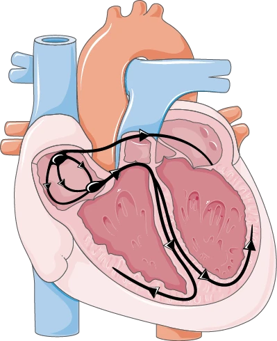
Anatomical study exhibit for the worked question below. Open the full-size view to inspect the structures.
Use the anatomical exhibit to work through the example. Choose an answer before opening the explanation.
Try it yourself
The illustration shows a normal frontal cutaway of the heart with the conduction pathways drawn as dark lines and directional arrows: activation spreads from the atria, converges at the junction between the atria and ventricles near the top of the interventricular septum, and then continues down the septum into both ventricles. This is a normal anatomical reference, not evidence of conduction disease. Two trainees debate conduction timing. Trainee 1 says the delay in the AV junction is what allows atrial contraction to complete ventricular filling before the QRS begins. Trainee 2 says delay in the bundle of His is the main source of the PR interval. Which assessment is correct? A. Trainee 2 is correct: the His bundle is the slowest conduction structure and produces most of the PR interval B. Trainee 1 is correct: AV junctional delay separates atrial depolarization from ventricular depolarization so atrial systole completes filling first C. Both are incorrect: the PR interval reflects only the speed of Purkinje fiber conduction D. Both are correct: the SA node creates the delay and both intervals correspond to atrial systole
Show answer and explanation
B. Trainee 1 is correct: AV junctional delay separates atrial depolarization from ventricular depolarization so atrial systole completes filling first
The AV junction is the deliberately slow-conduction region, and its delay electrically staggers atrial and ventricular depolarization so the atrial kick finishes filling the ventricles before systole begins with the QRS. The His bundle and Purkinje system conduct rapidly, so Trainee 2 misattributes the delay. In the illustration, the pathway converges at the AV junction at the top of the interventricular septum before the His bundle descends the septum; that anatomical sequence shows the two structures are distinct, and the schematic supplies their spatial relationship without indicating where the physiologic delay occurs, which is what the examinee must know.
Artifact or Arrhythmia? Verifying What You Are Looking At
Artifact is the most persistent look-alike in monitoring work. Muscle tremor, electrode movement, and baseline wander can imitate fibrillatory or wide-complex activity, so verify across leads and confirm beat-to-beat consistency before naming anything.
Worked scenario: a telemetry recording shows a chaotic, wandering baseline with an apparently irregularly irregular rhythm, and the first impulse is to describe atrial fibrillation. The better decision is to inspect the second lead on the same recording, where clear upright P waves precede every QRS at a constant PR interval. Measured with calipers, the QRS-to-QRS intervals are actually regular, and the apparent irregularity is baseline drift from electrode movement. The mistaken label matters because a recorded impression of atrial fibrillation can feed anticoagulation and workup decisions far upstream; the measurement sequence exposes it in seconds.
Train on the general verification cues. Artifact typically produces deflections wider or narrower than any physiologic QRS, signals that coincide with patient movement or cable handling, findings absent from a parallel lead, and patterns that leave no space where a T wave should sit. When artifact is the best explanation, document it as suspected artifact with your reason — movement noted, finding absent in lead two — rather than silently discarding the segment. An unexplained gap in a recording is its own documentation problem.
- Compare the suspect segment against a second lead on the same recording.
- Check whether deflection widths fall within plausible QRS dimensions.
- Correlate timing with patient movement, cable contact, or electrode problems.
- Verify regularity with a measurement tool, never a visual impression.
- Record suspected artifact with its supporting reason; never erase a segment unexplained.
Naming Tachycardias: Narrow, Wide, Regular, Irregular
QRS width and regularity split the tachycardias. Regular narrow-complex rhythms point toward supraventricular origins; wide-complex rhythms raise ventricular considerations. Naming requires measuring the QRS first, never inferring origin from rate alone.
Worked scenario: a strip shows a perfectly regular tachycardia near 180 beats per minute with no identifiable P waves, and the fast, unusual-looking trace tempts you to call it ventricular tachycardia. The better decision is to measure the QRS: in this example it is narrow, under 0.12 seconds, and the rhythm is regular. The defensible description is a regular narrow-complex tachycardia, P waves not identified, consistent with a supraventricular origin for description purposes, flagged for review. The distinction matters because the two families carry different immediate implications, and the downstream reviewer depends on your width measurement, not your adjective.
Respect the limits of the simplified exam frame. A wide-complex tachycardia can also represent a supraventricular rhythm with aberrant conduction, so a wide QRS raises a consideration rather than proving a mechanism, and definitive mechanism claims belong to the interpreting clinician. Paper scenarios are not universal real-world behavior. Your exam-day task and your workplace task are the same: measure QRS width and regularity accurately, describe what the measurements show, and flag rather than conclude when the picture is ambiguous.
AV Blocks: Reading the PR Interval Beat by Beat
Each AV block shows distinct PR-interval behavior: first-degree is consistently prolonged, Mobitz I lengthens and then drops a beat, Mobitz II drops without warning, and third-degree shows atrial and ventricular activity running independently.
Learn the blocks as PR-interval stories. First-degree AV block shows every P wave conducted with a constant PR interval beyond the usual upper limit — near 0.28 seconds in a typical worked example. Second-degree Mobitz I (Wenckebach) shows PR intervals lengthening across successive beats until a QRS drops, after which the cycle restarts short. Mobitz II shows constant PR intervals with sudden, unexpected dropped QRS complexes. Third-degree shows P waves and QRS complexes marching at unrelated rates with no fixed relationship. The table below turns these into side-by-side discriminators you can rehearse.
Mini-case: a strip looks like first-degree block, and the plausible mistake is measuring one PR interval and averaging the rest by eye. The better decision is to measure the PR before every beat and compare consecutive values. Here the intervals run 0.16, 0.24, 0.32 seconds before a dropped QRS, then reset — second-degree Mobitz I, not first-degree. It matters because the block categories differ in how reviewers prioritize them, and beat-by-beat comparison is the only measurement that separates a stable pattern from a progressive one.
| Pattern | Typical rate | Regularity | P waves | PR behavior | Key discriminator |
|---|---|---|---|---|---|
| Normal sinus rhythm | 60–100 | Regular | One per QRS, upright in lead II | Constant, normal duration | All measurements within usual ranges |
| Sinus bradycardia | Below 60 | Regular | One per QRS | Constant, normal duration | Sinus pattern at a slower rate |
| Atrial fibrillation | Variable | Irregularly irregular | Absent; fibrillatory baseline | Not measurable | No P waves plus irregular ventricular response |
| Atrial flutter | Often about 150 ventricular | Often regular | Flutter waves, sawtooth-like | Often a fixed conduction ratio | Atrial rate far exceeds ventricular rate |
| First-degree AV block | Usually 60–100 | Regular | One per QRS | Constantly prolonged | Every beat conducted, PR uniformly long |
| Second-degree Mobitz I | Variable | Grouped beating | Present | Lengthens then drops; resets | Progressive PR lengthening before the dropped beat |
| Second-degree Mobitz II | Variable | Irregular at drops | Present | Constant until sudden drop | Dropped QRS without PR change |
| Third-degree AV block | Ventricular escape rate | P and QRS each regular, independent | Present | No fixed PR relationship | Atrial and ventricular activity dissociated |
| Ventricular tachycardia pattern | Fast | Usually regular | Not identifiable | Not measurable | Wide, regular complex tachycardia |
Documentation and Escalation Language That Matches Technician Scope
Technician value lies in complete, neutral descriptions: what was measured, where it appeared, and what was flagged for review. CCI's Code of Ethics emphasizes patient privacy and ongoing competence, both of which surface directly in monitoring documentation.
Use a one-line descriptive template for every finding: rate, regularity, P waves, PR interval, QRS width, a rhythm name if the measurements support one, the time and duration of the event, and any artifact notes. Write what you measured, not what you suspect happened clinically. A phrase such as 'regular wide-complex tachycardia, 180 beats per minute, 42 seconds, P waves not identified — flagged for review' tells a reviewer everything needed, while 'possible VT, clinically concerning' crosses into interpretation that is not the technician's call.
Ground the professional habits in the code you will be held to. CCI's Code of Ethics requires credentialed professionals and candidates to protect patient privacy and confidentiality unless disclosure is required by law, to place patient safety above other interests, and to maintain competence through continuing assessment and education. In monitoring work those commitments are concrete: recordings and descriptions handled under facility privacy policy, no patient-identifying commentary in working notes, no silent deletion of data segments, and escalation through the defined channel when a finding needs review rather than through informal side conversations.
A Ten-Strip Drill, a Self-Check Rubric, and a Study Sequence
Run a ten-strip drill: apply the fixed sequence to each strip, write a one-line description, and score yourself against a rubric. Structure study as measurement drills first, artifact training second, then tachycardia and block differentials, then timed mixed sets.
The exercise: collect ten printed rhythm strips from a textbook or practice bank, allow about two minutes each, and score every strip against five checks — rate within ten percent of a verified count, regularity confirmed with at least two measured intervals, the P-to-QRS relationship stated, QRS width measured, and no diagnostic conclusion beyond what the measurements support. A reasonable learning milestone is eight of ten strips passing all five checks before you move to mixed timed drills. That score is a study milestone only, not a prediction of any exam result.
An adaptable sequence: spend the first stretch drilling the measurement sequence on sinus variants until descriptions are automatic; the second on artifact recognition using deliberately messy recordings; the third on the tachycardia split by QRS width; the fourth on the AV block table until each discriminator is automatic; and the final stretch on timed mixed sets with a written one-line description for every strip. Adjust the proportions to your rubric results — whichever check fails most gets the next block of practice. You can pair the drill with extra practice questions, then return to the broader study guide library to keep your routine consistent across materials.
Readiness checks before you stop drilling: you can produce a complete one-line description for an unfamiliar strip in about a minute; you can say, for any finding, whether you would describe it or flag it for review; you can name the PR behavior of all four AV blocks from memory; and you can list three artifact mimics together with the verification step that exposed each one. Re-run the rubric whenever you change study materials, so your scoring stays comparable.
References and further reading
Use these references to explore the concepts and check the latest information from the relevant organizations.