
Build your readiness for the NMTCB CNMT exam around reasoning with nuclear medicine concepts rather than memorizing isolated facts. The most useful approach is to study each radiopharmaceutical through its localization mechanism, then practice separating physiologic uptake, technical artifact, and pathology on paper cases. Pair that with a weekly image-review exercise, a decision table for common findings, and a self-check rubric so you can measure whether your differentials are getting faster and more complete. Administrative details such as eligibility and scheduling belong to the NMTCB itself; this article focuses on the reasoning the exam content draws on.
Why localization mechanisms, not drug lists, decide radiopharmaceutical questions
Learn each radiopharmaceutical by its localization mechanism first. Mechanisms explain expected distribution, justify artifact patterns, and let you predict behavior in altered physiology instead of memorizing a separate fact for every agent.
Compare two ways of knowing Tc-99m MAA. A list-based approach records that it images perfusion. A mechanism-based approach records capillary blockade: particles lodge temporarily in pulmonary capillary beds in proportion to flow. The mechanism predicts that markedly reduced flow produces reduced uptake, that excessive particle count shifts the blockade pattern, and that right-to-left shunting shows renal or cerebral activity. You can now answer questions you never specifically memorized.
Build your own mechanism table and force every agent you review through one category. When a practice vignette describes unexpected distribution, ask which mechanism the agent uses and what would disrupt it. A phagocytic agent behaving abnormally points to liver reticuloendothelial function; a receptor-binding agent behaving abnormally points to receptor saturation or blockade. The mechanism converts a surprising finding into a short, checkable list of causes.
- Capillary blockade (for example, Tc-99m MAA in lung perfusion imaging)
- Active transport or trapping (iodide in thyroid, MDP in bone by exchange at the hydroxyapatite surface)
- Phagocytosis (Tc-99m sulfur colloid in reticuloendothelial cells)
- Compartmental filling (blood pool agents, cerebrospinal fluid imaging)
- Receptor or antibody binding (targeted peptide and antibody agents)
- Simple diffusion or excretion (renally cleared tracers imaged during transit)
| Finding on the image | Physiologic explanation | Technical or procedural explanation | Pathologic explanation to keep in the differential |
|---|---|---|---|
| Focal uptake in the chest on a bone scan | Costal cartilage or injection-site activity in the arm | Contaminated glove or gurney transfer | Rib metastasis or fracture |
| Diffuse lung uptake on a bone scan | Known therapy-related finding documented for some agents | Radiopharmaceutical issue at preparation | Metastatic pulmonary calcification |
| Absent renal activity on a bone scan | Urinary diversion or pelvic kidney anatomy | Wrong acquisition field | Diffuse metastatic replacement documented in advanced disease |
| Stomach activity on an iodine scan | Normal physiologic iodide uptake | Residual contamination near the mouth | Ectopic gastric mucosa in the intended clinical context |
Separating artifact from pathology: the three-column habit
For any notable finding, write three candidate explanations: physiologic, technical or procedural, and pathologic. Rank them against the vignette. Practicing this habit builds the differential reasoning nuclear medicine demands and prevents premature pathologic calls.
The three-column habit works because nuclear medicine images rarely carry a single possible meaning. A cold defect in the liver on a sulfur colloid study could be a tumor, a cyst, a breast shadow attenuating counts, or a processing defect. The vignette clues that separate these are exactly the details scenario practice should train you to use: patient positioning, history of surgery, and whether the finding appears on multiple projections.
Practice the habit in writing. For ten anonymized teaching images, record the finding, the three columns, and the single observation that would confirm or eliminate each candidate. For example, repositioning the patient and re-acquiring eliminates attenuation and positioning causes; reviewing the injection log eliminates infiltration. When your written eliminations match the teaching file's final explanation, your reasoning chain, not just your answer, is sound.
Explore in 3D: Liver and Gallbladder
Locate the gallbladder on the inferior surface of the liver, then inspect the included bile duct. The liver and gallbladder are accessory digestive organs outside the food passage.
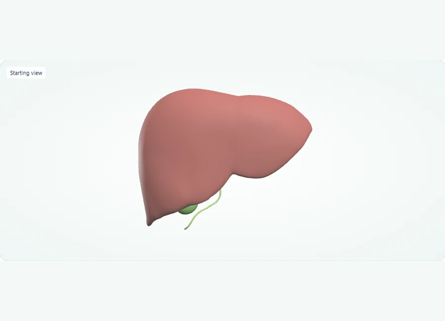
Rotate, isolate structures, and test your recall.
Rotate below the liver to find the gallbladder on its inferior surface.
Select the gallbladder and isolate it to inspect its shape. Use Show all before comparing its position with the liver.
Select and hide the liver, then select the bile duct to inspect its course. Complete hepatic and cystic duct junctions are not separately represented.
Try it yourself
Which accessory digestive organ lies on the inferior surface of the liver?
Show answer and explanation
Gallbladder
The gallbladder sits on the inferior (underside) surface of the liver and stores bile; bile leaves via the bile duct shown in this model.
- The gallbladder lies on the inferior surface of the liver.
- The liver and gallbladder are accessory organs — they are outside the food passage.
- The bile duct is included on this model, but the junctions between hepatic and cystic ducts are not shown or separately labelled.
Worked scenario 1: the hot chest finding on a bone scan
A vignette gives a focal area of increased uptake in the thorax of a bone scan. The plausible mistake in this scenario is calling metastasis immediately; the better decision is checking the injection history and the pattern of the uptake first.
Scenario: a patient injected in the left antecubital area shows focal uptake over the left anterior chest on anterior and oblique views, with faint linear uptake tracking toward the axilla. A first-pass reading calls this a left rib metastasis and moves on. The plausible mistake is ignoring the tracer pathway. Antecubital injection plus a linear axillary track strongly suggests radiopharmaceutical infiltration with lymphatic tracking, not bone pathology.
The better decision follows from evidence gathering already in the vignette: confirm the injection site matches the uptake, note whether the finding lies over bone or over soft tissue when compared with the opposite projection, and check whether the uptake appears in multiple views consistent with superficial tissue. Why it matters: infiltration is a procedural finding with quality implications, while a metastasis changes patient management. Recording both the image conclusion and the injection documentation is the complete answer.
Worked scenario 2: suppressed thyroid uptake and the medication history
When a thyroid uptake study shows low uptake, medication interference is a leading explanation. The plausible mistake in this scenario is diagnosing thyroiditis from the number alone; the better decision is reconstructing the patient's medication timeline before interpreting.
Scenario: a patient referred for a thyroid uptake measurement shows uptake well below the reference range for the technique. The first-pass reading attributes this to thyroiditis. The plausible mistake is treating the uptake value as a stand-alone result. Exogenous iodide from recent contrast studies or amiodarone, thyroid hormone therapy, and antithyroid medications all suppress uptake through different mechanisms, and the correct next step differs for each.
The better decision is to interrogate the timeline: ask about recent iodinated contrast, review thyroid hormone and antithyroid drug use with dates, and compare the uptake result with the patient's hormone levels. If the patient began levothyroxine weeks earlier, suppressed uptake reflects exogenous hormone suppression rather than primary thyroiditis. Why it matters: the differential changes the reported interpretation and the clinical recommendation. Tying the number to physiology and history, rather than to a memorized reference band, is the reasoning this subject calls for.
Radiation safety as a decision question, not a slogan
Study safety as trade-off reasoning: compare shielding, distance, and time options for a specific task and justify the choice. Paper scenarios let you practice selecting the option that reduces dose without compromising the study or the procedure.
For the NMTCB context, treat ALARA as a decision rule rather than a phrase to recall. Practice vignettes on dose administration for a pediatric patient, management of a radioactive spill, or handling of a compromised study after a misadministration is suspected. For each one, order the actions deliberately: protect people first, contain contamination, document, and notify according to your department's licensed procedures. Rehearse that ordering out loud until it is automatic.
Practice with written scenarios instead of improvised drills. Example: a dose is prepared for a study that must be rescheduled. Compare keeping the dose, reordering later, or adjusting the schedule within the radiopharmaceutical's usable window. The reasoning exercise — half-life, decay over the delay, institutional policy on dose acceptance — trains exactly this kind of comparison. Never rehearse these decisions with live sources outside supervised, licensed settings; the paper version captures the judgment you are building.
Telling the NMTCB credential family apart
The NMTCB maintains the general CNMT exam plus post-primary and specialty credentials. Do not study them interchangeably: each targets a different scope, and mixing their content wastes review time.
The NMTCB lists several distinct examinations: the general nuclear medicine exam leading to the CNMT credential, the post-primary NMTCB(CT) exam, the NCT and PET specialty examinations, and the Nuclear Medicine Advance Associate (NMAA) examination. Each targets a different scope of practice, and their content emphasis differs accordingly — CT fundamentals for the CT credential, PET-specific practice for PET. A useful exercise is to write one sentence describing the intended scope of each credential from the NMTCB applicant pages before you open a single study chapter.
Check which credential your goal requires before building a plan. If you are sitting for the general exam, a PET physics chapter borrowed from a specialty syllabus may exceed what you need, while omitting general planar and SPECT topics would leave gaps. The NMTCB's own applicant materials identify preparation resources per examination, so map your syllabus to the specific credential name, not to 'nuclear medicine' generally.
A four-week preparation sequence with a self-check rubric
Organize review as mechanism study, artifact drills, scenario practice, then mixed timed sets. Score yourself each week against a rubric so readiness is measured by reasoning quality, not by how many pages you covered.
A workable adaptable sequence: in week one, build the mechanism table for every agent in your syllabus and explain each to a peer without notes. In week two, run the three-column artifact exercise on ten teaching images and log your eliminations. In week three, write out worked scenarios — one hot finding, one suppressed-uptake case, one safety ordering task — and time yourself. In week four, mix domains in timed sets and use the free practice questions on this site to surface weak areas, then loop back to the mechanism table for anything you missed.
Use this rubric weekly. For each practice scenario, award yourself: one point if you produced three candidate explanations before answering; one point if your eliminated causes cited an observation from the vignette rather than a guess; one point if you named the localization mechanism involved; one point if you identified the procedural or documentation step that follows. A rising score across weeks is a learning milestone showing your reasoning is consolidating — it is a study signal, not a prediction of your exam result.
References and further reading
Use these references to explore the concepts and check the latest information from the relevant organizations.