
Organize ABEM review around diagnostic discriminators: for each chief complaint, learn the worst-first differential, the one feature that separates the leading diagnosis from its closest mimic, and the escalation trigger when the patient does not respond as expected. Test yourself with timed case scenarios and score the reasoning steps, not only the final answer.
Why a Topic Checklist Fails: Build Discriminator Maps Instead
ABEM review pays off when organized around discriminating features that separate look-alike emergencies, because emergency care rewards rapid branching decisions rather than exhaustive textbook knowledge recalled in textbook order.
A discriminator is the specific history item, examination finding, or early test result that changes your working diagnosis. Classic presentations such as crushing substernal chest pain are easy to memorize; the cognitive work lies in the patient with indigestion-like discomfort whose real problem is dissection, embolism, or esophageal rupture. Build a one-page map for each chief complaint that lists the top three mimics and, next to each, the discriminator that rules it in or out.
Layer worst-first reasoning on top of that map. Rank the differential partly by how quickly a missed diagnosis harms the patient, not solely by how common it is. Aortic dissection sits high on the chest-pain map even though it is uncommon, because hours matter. When you study any topic, ask two questions: what is the most dangerous thing this could be, and what single observation would make me change course? Write both answers into your notes.
Chest Pain: Separating ACS From the Mimics That Punish the Wrong Treatment
Chest pain review should pair each leading diagnosis with its closest mimic and one discriminating finding, so management rests on explicit reasoning about the differential instead of pattern matching to a stereotype.
Worked scenario: a 58-year-old man presents with sweating and vague epigastric discomfort. The initial instinct is acute coronary syndrome, and anticoagulation is drawn up. On examination, his right arm pressure is markedly lower than his left, the pain began suddenly and radiates to the back, and the chest radiograph suggests a widened mediastinum. The better decision is to pause anticoagulation, reassess pulses and blood pressure in both arms, obtain bedside echocardiography, and proceed to CT angiography. This matters because anticoagulating a dissection can convert a survivable problem into a fatal one.
The reusable lesson is that every ACS presentation deserves a sixty-second mimic screen: characteristics of the pain, bilateral blood pressures, a look for pulse deficits, and a mediastinum check on the film. Do the same pairing for the other common causes you review — pericarditis versus ST-elevation patterns, pulmonary embolism versus anginal-equivalent dyspnea, and esophageal rupture versus reflux after vomiting. Each pair should end with a sentence you can recite: what feature moves the diagnosis from one column to the other.
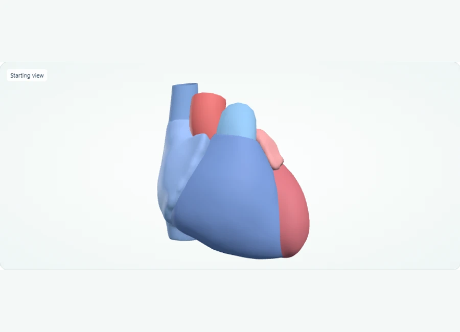
Explore in 3D: Heart chambers and great vessels
A four-chambered heart viewed from the front. The right ventricle supplies the pulmonary circulation via the pulmonary trunk; the left ventricle supplies the systemic circulation via the ascending aorta. This model shows external chamber surfaces and the visible great vessels only — no internal valves or conduction system.
Rotate, isolate structures, and test your recall.
Choose Front and compare the four chambers. The right ventricle forms most of the anterior surface; the left ventricle contributes to the apex.
Select and isolate each chamber in turn, then use Show all to restore its relationship to the surrounding structures.
Select the pulmonary trunk and ascending aorta in turn. Name the circulation supplied by each, then use Test recall to practise identifying the listed structures.
Try it yourself
Which ventricle supplies the pulmonary trunk, and which supplies the ascending aorta?
Show answer and explanation
The right ventricle; it serves the pulmonary circulation.
On the front view, the right ventricle is the anterior-most chamber, and its outflow tract continues as the pulmonary trunk carrying blood to the lungs. The left ventricle instead empties into the ascending aorta for the systemic circulation.
- The right ventricle pumps blood into the pulmonary trunk toward the lungs; the left ventricle pumps into the ascending aorta to the body.
- The superior vena cava returns blood to the right atrium from above the heart.
- The two atria sit superior to the two ventricles; atria receive blood, ventricles eject it.
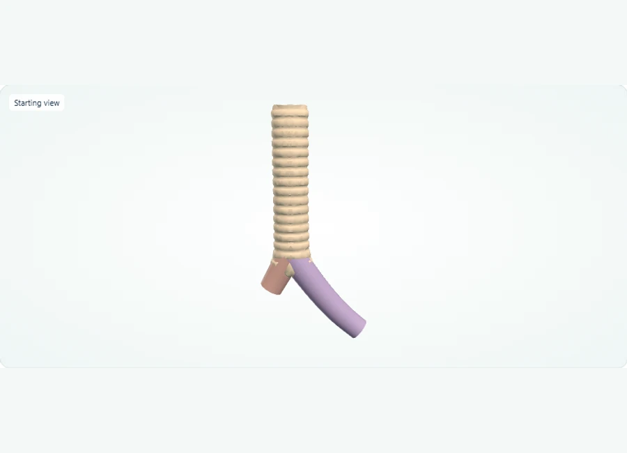
Explore in 3D: Trachea and Main Bronchi
This model shows the trachea and its division into the right and left main bronchi. Only these three airway structures are present: there are no lobar branches, alveoli, or other respiratory structures, and the model shows normal anatomy with no pathology or devices.
Rotate, isolate structures, and test your recall.
Set the view to front and zoom to see the whole model: the trachea is the single central tube, with the right main bronchus and left main bronchus branching from its lower end.
Select each of the three structures in turn (Trachea, Right main bronchus, Left main bronchus), using isolate to view one at a time and show all to restore the full model.
Use Test recall to hide the labels; when a structure is highlighted, name it before pressing Reveal name, then use Return to study to continue.
Try it yourself
Test recall highlights the structure branching from the lower end of the trachea toward the body's left side. Which structure is it?
Show answer and explanation
Left main bronchus
In the front view, the trachea runs down the midline and splits into two main bronchi. The branch passing to the viewer's right (the body's left side) is the left main bronchus, while the branch to the body's right side is the right main bronchus.
- The trachea is the central airway that divides into two main bronchi, one for each lung side.
- The right main bronchus and left main bronchus are the first branches off the trachea; this model contains no smaller (lobar) branches or alveoli beyond them.
- Because only three structures are selectable, identification practice here means telling the trachea from the right versus left main bronchus.
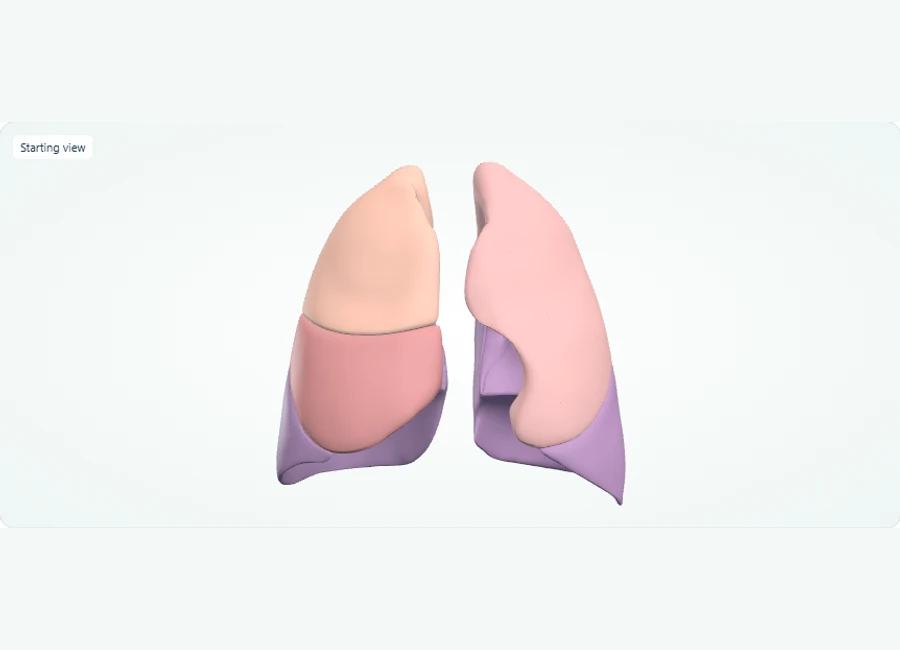
Explore in 3D: Lung lobes
This model shows the five lung lobes: three on the right (superior, middle, inferior) and two on the left (superior, inferior). The left lung has no middle lobe. Each colored lobe is a distinct selectable structure, separate from segments and bronchial branches. As a normal surface model, it shows healthy anatomy only.
Rotate, isolate structures, and test your recall.
Choose Front. Use the Structure list to select each lobe and count three on the anatomical right and two on the left.
Select and isolate the right middle lobe, then use Show all and Back to inspect how it relates to the other right lung lobes.
Start Test recall, name the highlighted lobe, then select Reveal name to check your answer.
Try it yourself
How many lobes does each lung have, and which lobe name occurs only on the right?
Show answer and explanation
The right lung has superior, middle and inferior lobes. The left lung has superior and inferior lobes, with no middle lobe.
A lobe is a gross anatomical division of a lung. The right lung has three lobes and the left has two. These surface models show lobes, not individual bronchopulmonary segments or surgical margins.
- The right lung has three lobes: superior, middle, and inferior.
- The left lung has only two lobes: superior and inferior; there is no left middle lobe.
- Lobes are distinct from segments and bronchial branches, and a normal model shows no tumors or surgical margins.
Visual anatomy review: Follow the digestive route and accessory organs
Distinguish the gallbladder from the connected biliary drainage pathway when reviewing the case.
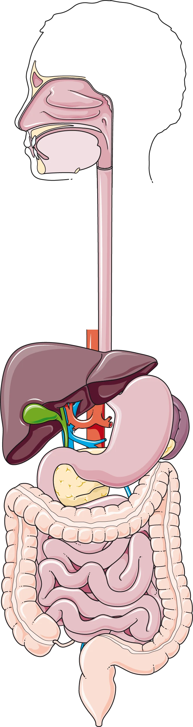
The digestive tract is a continuous passage; the liver, gallbladder and pancreas are accessory organs. Open the full-size illustration for the smaller abdominal structures.
Trace the esophagus into the stomach and then the small intestine. The duodenum comes first, followed by the jejunum and ileum. The gallbladder lies beneath the liver; it is not part of the passage through which food travels.
In Z-Anatomy, locate the stomach, duodenum, liver and gallbladder. Rotate the visceral structures to inspect their relationships, then describe the food passage without including the accessory organs in it.
Try it yourself
An upper endoscope passes through the esophagus and stomach. Which small-intestinal segment does it reach first, and is the gallbladder part of that route?
Show answer and explanation
It reaches the duodenum first. The gallbladder is an accessory organ outside that food passage.
Separate the route of access from the target of a procedure. The diagram supports location recall; the actual operative findings and work performed determine the procedure being described.
The Dyspneic Patient: One Table to Separate Pump, Pipe, and Perfusion Problems
Dyspnea assessment hinges on separating pump failure, airway obstruction, perfusion loss, and chest-wall problems, because each responds to different immediate therapy and the wrong first move can worsen the patient.
A useful organizing frame sorts dyspnea into pump problems (heart failure, ischemia), pipe problems (asthma, COPD, airway obstruction), perfusion problems (pulmonary embolism, severe anemia), and container problems (pneumothorax, effusion). Physical examination discriminators do real work here: neck vein distension and peripheral edema point toward the pump; prolonged expiratory wheeze points toward the pipe; acute unilateral silence with tracheal shift points toward a tension pneumothorax; clear lungs with unexplained hypoxia raises the perfusion question.
Treat this table as a teaching simplification rather than a decision rule. Real patients combine categories, and imaging and laboratory confirmation are required before definitive therapy. The value of the frame is that it forces you to verbalize which category you believe and what evidence would move you. When you review a dyspnea case, state the category, the discriminator that supported it, the immediate action, and the finding that would make you abandon that category. Practicing that sentence structure is more transferable than rereading any single disease page.
| Category | Typical discriminators | Immediate bedside focus | Hazard if confused |
|---|---|---|---|
| Pump (heart failure) | Orthopnea, elevated neck veins, crackles, edema | Position, oxygen, afterload and fluid balance thinking | Giving a large fluid bolus to a wet patient |
| Pipe (asthma/COPD) | Wheeze, prolonged expiration, known reactive disease | Inhaled bronchodilation, assessment of work of breathing | Missing a pneumothorax hiding behind wheeze |
| Perfusion (PE, anemia) | Clear lungs with hypoxia, pleuritic pain, risk factors | Risk stratification and definitive imaging pathway | Reassuring labs of infection while clot progresses |
| Container (pneumothorax) | Unilateral decreased breath sounds, acute pleuritic onset | Decompression if tension physiology appears | Waiting on imaging while the patient deteriorates |
Undifferentiated Shock: Sepsis and the Mimics That Change the Fluid Plan
Reviewing shock means pairing sepsis with its mimics — hemorrhage, adrenal crisis, embolism, anaphylaxis — and rehearsing what changes when a presumed septic patient fails to respond as expected.
Worked scenario: an 82-year-old woman arrives confused and hypotensive with a low-grade fever. The working label becomes urosepsis, and fluid resuscitation proceeds without reassessment. She remains hypotensive after several boluses, her abdomen is distended and tender, and her hemoglobin result returns low. The better decision is to treat this as unexplained shock: consider hemorrhage and adrenal insufficiency alongside infection, order cross-matched blood, and add corticosteroid coverage when adrenal crisis remains plausible. This matters because continuing boluses into an occult hemorrhage deepens the problem, while sepsis pathways assumed to be working can delay the correct intervention.
Two named concepts make this scenario teachable. First, shock categories — distributive, cardiogenic, hypovolemic, obstructive — predict different responses to the same treatment. Second, reassessment is a diagnostic act: a patient who fails to respond to your initial therapy has given you information, and the differential must widen rather than the dose simply increase. In your notes, attach a written trigger to every sepsis-style pathway: the specific observation that halts the pathway and reopens the differential.
Neurologic emergencies: The Five Mimics That Routinely Imitate Stroke
Neurologic review should center on the short list of stroke mimics and their discriminators, because bedside glucose, seizure history, and facial-weakness pattern each redirect a time-critical pathway.
Build a five-item mimic list and learn one discriminator per item. Hypoglycemia: a bedside glucose value can fully explain a dense hemiparesis and is checked within the first minutes. Seizure with Todd's paresis: the weakness follows a witnessed or suspected seizure and typically improves over hours, unlike a fixed stroke deficit. Complicated migraine: a spreading aura and prior similar episodes shift the picture. Peripheral Bell's palsy versus a central lesion: forehead involvement and the absence of other deficits suggest a peripheral cause, while forehead sparing, arm weakness, or dysarthria point centrally. Conversion or functional presentations: an internally consistent, incongruent examination without vascular risk factors invites careful reassessment rather than dismissal.
The practical drill is a habit, not a fact list. Before any neuro-imaging pathway, the checklist order is glucose first, seizure history second, onset and progression third, and a structured cranial-nerve screen fourth. Rehearse saying the exclusion phrase aloud: a stroke diagnosis requires that the mimic list has actually been worked through, not merely assumed absent. Practice on written cases where the vignette deliberately buries the glucose value or the seizure witness in the middle of the narrative, because that is where a rushed reader drops it.
Capacity, Consent, and Refusal: Documenting Decisions in the Emergency Setting
Ethics review should treat capacity assessment, consent, and refusal documentation as applied clinical reasoning skills, since emergency settings routinely demand decisions with incomplete information and unaccompanied patients.
Distinguish capacity from competency: capacity is a clinical judgment about a specific decision at a specific moment, while competency is generally a legal determination. Assess capacity against its commonly described elements — the patient's ability to understand the relevant information, appreciate that it applies to their own situation, reason through the options, and communicate a stable choice. Note that capacity is decision-specific: a patient may retain capacity to refuse a low-risk intervention while lacking it for a high-risk one.
When a patient with capacity refuses a recommended intervention, the documentation task is to record the elements above, the risks discussed, the alternatives offered, and the opportunity for questions — the reasoning, not a single signature line. Pair this with the parallel skill of medical decision-making documentation: recording why the chosen pathway was selected over plausible alternatives, which is the written version of the discriminator reasoning taught throughout this guide. Study ethics as short written cases: draft the refusal note, then check it against the four capacity elements and confirm each one is visible on the page.
A Six-Week Sequence and a Rubric That Scores Reasoning, Not Recall
A workable sequence moves from organ-system discriminator maps, through timed mixed case sets, to self-scoring against a rubric that grades the reasoning steps rather than the final answer alone.
Suggested adaptable sequence: weeks one and two, build discriminator maps for eight to ten high-priority chief complaints and review each map aloud until the mimic pairs and discriminators come without notes. Weeks three and four, work timed case sets from your practice-question resources, writing a one-line differential and one-line next step before reading the explanation. Weeks five and six, mix domains and add documentation drills: one capacity-and-refusal note and one decision-making note per session.
Practical exercise with a self-check rubric: pick ten chief complaints and, for each, produce the top three differentials, one discriminator per differential, and one escalation trigger — target completing all ten within a fixed time you set yourself. Score one point per completed item and review anything scoring below your own milestone; these scores track learning progress and do not predict exam outcomes. Readiness checks before sitting down to intensive review: you can recite three mimics per major complaint from memory; you can name the observation that would stop each major pathway; and your written case notes state a rationale, not just a diagnosis.
One short administrative note: for certification requirements, examination formats, and eligibility details, rely on the issuer directly at abem.org rather than secondary summaries.
References and further reading
Use these references to explore the concepts and check the latest information from the relevant organizations.