
Study for ABU urology certification by organizing content into paired concepts, rehearsing case-based decisions on paper, and practicing structured documentation. For eligibility, format, and scheduling details, rely on the American Board of Urology directly at abu.org rather than any third-party summary.
Mapping Urology Into Paired Concepts Instead of Topic Lists
Build your core review around contrasting concept pairs, because case-style practice asks you to choose between two similar entities, and drilling each contrast builds exactly that discrimination.
Start with four pairs that organize nearly the whole field: upper versus lower urinary tract, benign versus malignant disease, functional versus structural problems, and pediatric versus adult presentations. For each pair, write one sentence explaining why the distinction changes management. For example, upper tract versus lower tract changes both the imaging you choose and the anatomical explanation of symptoms, so the pair is a decision tool, not a memorization label.
Then attach each named condition to exactly one slot in this map. Testicular torsion is acute, structural, and time-sensitive; benign prostatic hyperplasia is chronic, structural, and lower tract; overactive bladder is functional and lower tract. When a condition fits two slots ambiguously, flag it for extra scenario practice, because ambiguity in your own map is where case reasoning stalls. A one-page hand-drawn map you can reproduce from memory is a stronger milestone than a completed flashcard deck, since reconstruction proves the structure is yours.
- Upper vs lower tract: changes imaging choice and symptom explanation
- Benign vs malignant: changes urgency, workup depth, and counseling
- Functional vs structural: changes whether you treat behavior, medication, or anatomy
- Pediatric vs adult: changes both differential lists and communication approach
Interpreting Urinalysis, PSA, and Imaging Without Overreading
Treat every laboratory value and image as a clue inside a context, never as a diagnosis. Practice stating, out loud, what each finding supports, what it rules out, and what it cannot address at all.
For urinalysis, deliberately separate three questions: is there blood, is there infection, and is there protein or casts? Make this separation concrete with a column drill: write the three questions as column headers, then re-read ten practice urinalysis reports and enter each result in the column it bears on, leaving blanks where a result says nothing. Hematuria without nitrites or leukocyte esterase belongs in the blood column and points away from infection toward a structural or oncologic question. If you cannot state the next confirmatory step for a result, you have recognized the finding but not interpreted it.
For prostate-related labs and imaging, apply the same discipline. A PSA value is a risk indicator influenced by prostate volume, inflammation, and recent instrumentation, not a cancer verdict; standard teaching emphasizes interpreting it alongside examination findings and history rather than in isolation. For imaging, practice selecting modality by question asked: a non-contrast study answers stone questions well, contrast-based upper tract imaging answers filling-defect questions, and ultrasound answers many scrotal and bladder-volume questions. Writing one line per modality about what it can and cannot show prevents the reasoning error of demanding more from an image than it provides.
Explore in 3D: Kidneys, Ureters and Bladder: The Urine Transport Pathway
This model shows five selectable structures: the right and left kidneys, the right and left ureters, and the urinary bladder. It demonstrates the gross pathway of urine transport from where urine forms (kidneys) to where it is stored (bladder). Note: the urethra and internal renal anatomy are not included in this model, so the pathway ends at the bladder.
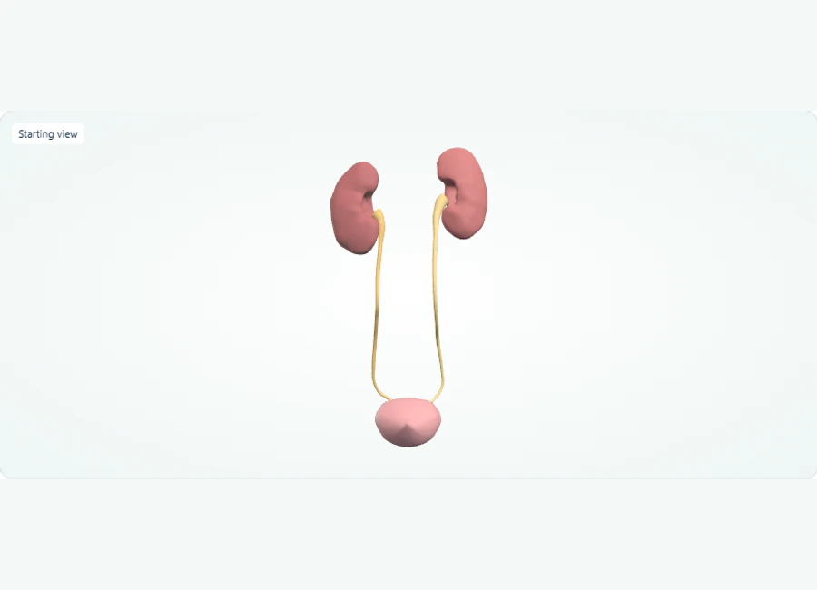
Rotate, isolate structures, and test your recall.
Choose Front. Select the right kidney, then its ureter, and follow the route toward the bladder with all structures visible.
Repeat on the left side. Select and hide each kidney separately if you want a clearer view of the ureters; use Show all to restore them.
Select the urinary bladder. Rotate the model and identify where the paired ureters meet the single bladder.
Try it yourself
Trace the three structures in the right-sided urine transport route shown here. What does the final structure do?
Show answer and explanation
Right kidney, right ureter, urinary bladder. The bladder stores urine until it leaves the body. (The left side follows the same pattern: left kidney and left ureter also deliver urine to the bladder.)
Urine flows from each kidney into its own ureter, which transports it down to the urinary bladder for storage. Both the right and left ureters empty into the single bladder. Because the urethra is absent from this model, the bladder is the terminal structure you can trace.
- Urine is formed in the kidneys; from each kidney, urine drains into that side's ureter (right ureter from right kidney, left ureter from left kidney).
- Each ureter carries urine downward to the urinary bladder, which stores urine until it is expelled; this model shows both ureters joining the bladder.
- This model is a gross transport overview only: the urethra and internal kidney structures are not shown, so urine flow beyond the bladder cannot be traced here.
Visual anatomy review: Trace the urinary tract
Use the normal urinary route to organize the location of leakage or obstruction described in the case.
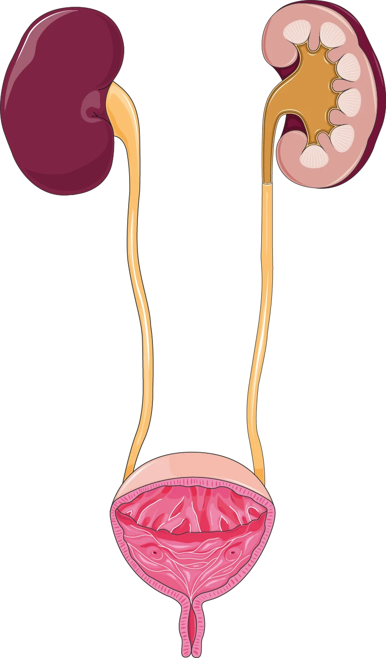
Follow each ureter from a kidney to the bladder; the urethra is the outlet below the bladder. This is a normal female anatomy reference, not an image of the patient.
The ureter carries urine from a kidney to the bladder. The urethra carries urine out of the bladder. Keep these names separate when reading an endoscopic route or locating an obstruction.
In Z-Anatomy, locate a kidney, its ureter and the bladder. Rotate the model, then trace the same route without the labels. Name the structure before and after the bladder separately.
Try it yourself
A scope enters the bladder through the urethra, then advances toward a kidney. Which tube does it enter after the bladder?
Show answer and explanation
The ureter on the side being examined.
Working backward along urine flow distinguishes ureteroscopy from inspection limited to the bladder. An anatomy diagram does not establish which procedure was completed.
Worked Scenario 1: The Hematuria Ordering Trap
Hematuria scenarios reward a two-step habit: first confirm and characterize the finding, then evaluate both upper and lower tracts. The instructive error is stopping after one reassuring result.
Scenario: a 58-year-old current smoker is found to have microscopic hematuria on an insurance screening dipstick. There are no urinary symptoms. The plausible mistake here is treating it as a transient benign finding, or alternatively ordering a single study, seeing a normal result, and closing the case. Both errors share one root: treating one negative or one positive data point as the whole answer instead of confirming the finding and then reasoning about anatomy.
The better decision follows a structured path: repeat the urinalysis to confirm persistent hematuria, take a focused history for trauma, infection, instrumentation, and family history of urothelial cancer, and then plan an evaluation that addresses both the kidneys and upper tracts and the bladder itself, because a normal upper tract image says nothing about the bladder. In a clearly labeled simplified teaching version of this case, note that smoking and age raise pre-test concern for urothelial malignancy, which is exactly why a negative single test should lower your vigilance only slightly. Practicing this scenario trains the habit of matching your remaining uncertainty to your remaining workup, and that habit is what makes a case write-up defensible on its own terms.
Worked Scenario 2: The Acute Scrotum and the Delay Decision
Time-sensitive urologic emergencies test sequencing, not diagnosis. The key learning point is recognizing when your suspicion level itself should change your actions before any test result returns.
Scenario: a 15-year-old presents with two hours of sudden severe left scrotal pain, nausea, and a high-riding, exquisitely tender testis with an absent cremasteric reflex on examination. The plausible mistake is treating ultrasound as a prerequisite gate: sending the patient to imaging with a long queue, and letting the referral process consume the window in which testicular viability is best preserved. The error is procedural sequencing, not knowledge of torsion itself.
The better decision, consistent with standard emergency teaching, is that when clinical suspicion of torsion is high from the history and examination, immediate urologic consultation and readiness for surgical exploration take priority over waiting for definitive imaging; imaging is for cases where suspicion is genuinely uncertain. Contrast this with a low-suspicion presentation, such as gradual onset pain with an intact reflex and a tender epididymis, where ultrasound to distinguish epididymitis from other causes is reasonable and antibiotics directed at likely pathogens follow. Write both branches out side by side. For every urologic emergency you study, record which branch point changes the order of actions; that recorded branch point becomes a concrete, reusable self-test.
Procedures and Documentation That Hold Up Under Scrutiny
Procedure review should pair the mechanical steps of common urologic procedures with the documentation that justifies them. Practice writing notes structured as indication, consent, findings, technique, and complications.
For each commonly tested procedure family, including urethral catheterization, cystoscopy, transurethral resection, and stent placement, learn three layers: the indication (why this patient needs this now), the key technical and sterile precautions, and the recognized complications with their early signs. A catheter placed for retention has a different justification and different documentation than one placed for output monitoring, even though the technique is identical. Layered learning like this keeps reasons attached to steps rather than letting the steps float alone.
Now practice documentation as a written exercise. Take a paper case of a transurethral resection and draft an operative note covering the indication and preoperative discussion, the findings at cystoscopy, the resection technique and landmarks, specimen handling, and any intraoperative events. Then audit your own note against a five-point rubric: could a colleague reconstruct the indication, would the consent discussion be evident, are findings specific rather than generic, is the technique described in sequence, and are complications or their absence explicitly stated? Notes that fail the audit usually reveal gaps in your underlying understanding of the procedure, which makes this documentation drill a genuine knowledge check rather than a writing chore.
Case Analysis: A Decision Table for Lower Urinary Tract Symptoms
Use a comparison table to force differential reasoning. Lower urinary tract symptoms in an older man illustrate how overlapping complaints hide different diseases with different next steps.
Case vignettes reward a fixed parsing routine: identify the patient and timeframe, list the findings, map each finding to the differentials it supports or weakens, and only then choose a next step. Practicing this routine on one rich clinical area, such as lower urinary tract symptoms, transfers to every other area because the routine, not the content, is what you are rehearsing.
The table below contrasts four common explanations for lower urinary tract symptoms in adult men. Use it two ways: cover the right-hand columns and generate them from the left, then reverse the exercise and generate the left column from the findings. Both directions should feel automatic before you consider this content reviewed.
| Condition | Distinguishing features | Typical next step in evaluation |
|---|---|---|
| Benign prostatic hyperplasia | Gradual obstructive and irritative symptoms, enlarged prostate on examination, no systemic signs | Symptom assessment, examination, and evaluation of kidney function before therapy |
| Prostate malignancy concern | May be asymptomatic; suspicious examination findings or elevated risk context rather than voiding symptoms alone | Risk discussion with the patient and further prostate evaluation when indicated |
| Urinary tract infection / prostatitis | Dysuria, urgency, possible fever or perineal pain; abnormal urinalysis | Urinalysis and culture, then targeted antimicrobial treatment |
| Overactive bladder (functional) | Urgency and frequency without obstruction or infection findings | Behavioral measures and fluid review before or alongside medication |
Ethics, Safety, and an Adaptable Preparation Sequence
Weave consent, shared decision-making, and safety habits into every scenario you study, then follow a repeatable four-phase sequence that ends with self-scored readiness checks rather than vague confidence.
Ethics in urology review is most durable when attached to cases rather than studied as isolated principles. For each worked scenario, add one line on informed consent: what were the material risks discussed, what alternatives were offered, and what would make a patient's choice valid? Add one line on safety: sterile technique for instrumentation, correct patient and side verification for any lateralized procedure, and clear escalation when a patient deteriorates. These additions cost seconds and turn every case into ethics and safety practice simultaneously.
An adaptable sequence: weeks one and two, build the paired-concept map from section one and self-test by redrawing it. Weeks three and four, work through lab and imaging interpretation drills, verbalizing finding-category-next-step chains. Weeks five and six, write out ten paper scenarios including the two patterns modeled above, each with an explicit decision, an expected mistake, and a consent and safety line. Final phase, complete timed mixed-question practice from your question bank and score case write-ups against the rubric below. Readiness checks you can trust: you can reproduce the concept map unaided, you can state a next step for any lab finding you identify, and your last three self-scored case write-ups meet every rubric line.
- Rubric line 1: Confirmed the finding before acting on it (hematuria-type cases)
- Rubric line 2: Evaluation addressed all relevant anatomic regions, not just one
- Rubric line 3: Emergency sequencing reflected suspicion level, not test availability
- Rubric line 4: Note or plan states indication, consent, findings, technique, complications
- Rubric line 5: Next steps matched the remaining uncertainty rather than defaulting to maximal testing
References and further reading
Use these references to explore the concepts and check the latest information from the relevant organizations.