
Prepare for ABO-style content by studying diagnoses in contrast pairs rather than as isolated topics: glaucoma versus other optic neuropathies, papilledema versus pseudopapilledema, retinal versus neurological field defects. Work through transposition and vergence calculations by hand, analyze case vignettes using a fixed reasoning template, and track your progress with a rubric instead of a page count.
Building a Study Architecture Around the Core Ophthalmology Domains
Organize preparation around the major clinical domains that define ophthalmology as a specialty: optics and refraction, glaucoma, retina, cornea and external disease, neuro-ophthalmology, pediatric ophthalmology and strabismus, oculoplastics, uveitis, and ophthalmic pathology.
A domain map works best when each domain is defined by its characteristic decision, not just its anatomy. Optics is about calculating and correcting; glaucoma is about separating pressure-related damage from mimics; neuro-ophthalmology is about localizing lesions anatomically. Write one sentence like this for every domain, then attach your study of each disease to that decision rather than to a list of facts.
Cross-domain linkages deserve their own study pass. Conditions such as diabetes and thyroid eye disease appear in retina, cornea, neuro-ophthalmology, and oculoplastics at the same time. Build a one-page map for each systemic disease with a column for every domain it touches: how it presents there, which tests that domain relies on, and which management logic applies. Practicing retrieval from that map weekly turns scattered facts into a network you can search under time pressure.
- Domain decision: optics = calculate; glaucoma = differentiate; neuro-ophthalmology = localize
- Systemic disease map: one page per disease, columns for each domain it affects
- Weekly rotation: touch every domain at least once per two-week cycle so nothing goes cold
Optics and Refraction: Transposition and Vergence Without Guesswork
Optics rewards exact procedure. Master transposition of spherocylindrical prescriptions, cylinder axis notation, vergence and power calculations, and prism basics as repeatable algorithms, then verify each result with a physical sanity check rather than intuition.
Vergence questions reward dimensional discipline. If an object sits 50 cm from a thin lens, the object vergence is −100/50 = −2.00 D; dropping the negative sign for real objects, or mixing centimeters with diopters, produces answers off by magnitude or sign. The better decision is to write the units on every line: distance in meters, vergence in diopters, sign convention stated once at the top of the page. Practicing with this written scaffolding on ten problems teaches you where your own errors concentrate — sign, units, or arithmetic — which is far more useful than doing thirty unexamined problems.
A sanity check catches transposition errors instantly: in any valid spherocylindrical prescription, the sphere value must equal one of the two principal powers and sphere-plus-cylinder must equal the other, in both notations. Checking +1.00 +1.00 × 90 against the original +2.00 −1.00 × 180 confirms the principal powers (+2.00 and +1.00) match before and after. Build this check into every optics problem you do, so it fires automatically during the exam.
- Transposition algorithm: add, flip sign, rotate axis 90° — all three steps, every time
- Sanity check: principal powers must match across notations
- Vergence discipline: meters, diopters, and sign convention written explicitly
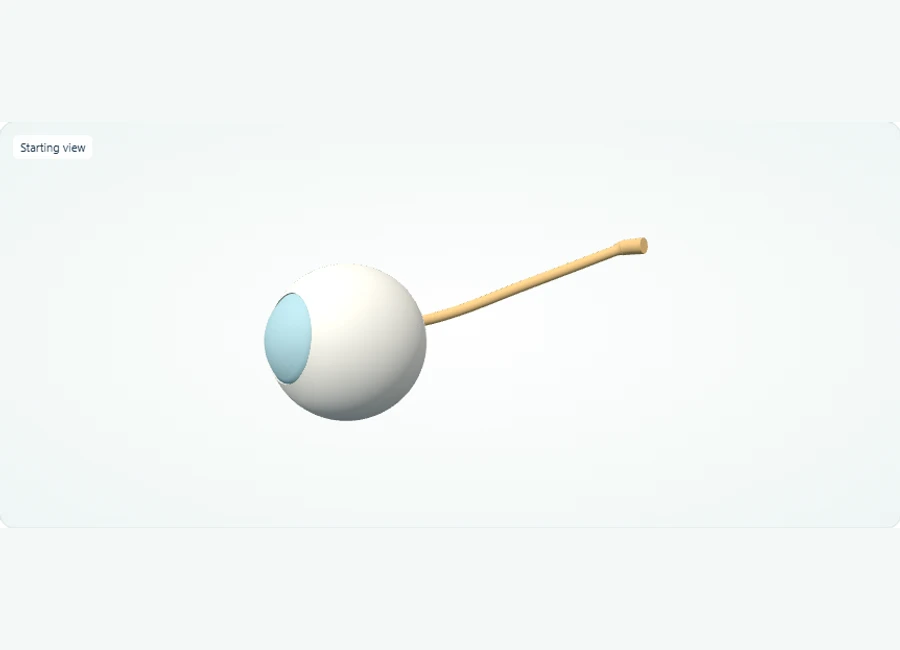
Explore in 3D: Right eye: layers, lens, and optic nerve
This model shows the right eye as five selectable parts: cornea, lens, retina, sclera, and optic nerve. The cornea and lens sit anteriorly, the retina lines the posterior interior, and the optic nerve exits posteriorly. Surfaces are opaque until hidden, so hiding the sclera is the key step to see inside.
Rotate, isolate structures, and test your recall.
Select and isolate the right cornea, then the right lens, to inspect these anterior structures individually. The study model uses opaque surfaces.
Use Show all. Select and hide the sclera to expose deeper layers, then isolate the retina to inspect its shape.
Use Show all and choose Side or Back. Select the optic nerve to trace its exit from the posterior eye.
Try it yourself
Which structure exits the posterior eye, and which light-sensitive layer lines the posterior interior?
Show answer and explanation
The optic nerve (cranial nerve II) exits posteriorly; the retina lines the posterior interior.
From the back of the model, the nerve trunk is the structure leaving the eyeball, and the retinal lining sits within the posterior interior. Note the entire retina is shown as one part, and the macula is not separately labelled in this model.
- Refractive structures (cornea and lens) are anterior; the light-sensitive retina is posterior.
- The optic nerve (cranial nerve II) leaves the eye posteriorly, carrying visual information toward the brain.
- The sclera is the opaque outer coat; hide it to expose the cornea-lens axis and the internal retinal lining.
Visual anatomy review: Separate anterior eye structures from the retina
Locate the posterior retina while keeping actual disease findings in the case description.
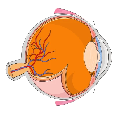
Normal eye cross-section for orientation. This is not an OCT scan or fundus photograph and does not depict a retinal tear, edema or retinopathy.
The cornea and lens are toward the front of the eye. The retina lines the posterior inner wall. Distinguish the structure examined or treated from the technique used to examine or treat it.
In Z-Anatomy, locate the eyeball and optic nerve and compare front and back views. Describe where the cornea, lens and retina belong before returning to the clinical or coding scenario.
Try it yourself
Would imaging the anterior segment and imaging the macular region examine the same anatomical location?
Show answer and explanation
No. The anterior segment is at the front of the eye; the macula is a region of the retina at the back.
Location matters even when an imaging technique has the same name. This normal schematic supports anatomy recall, not a diagnosis from an actual clinical image.
Glaucoma Versus Other Optic Neuropathies: Differentiating the Disc and the Field
Glaucoma study should center on differentiation: glaucomatous cupping versus non-glaucomatous optic neuropathy, open-angle versus angle-closure mechanisms, and pressure findings that must be interpreted alongside disc and field evidence rather than in isolation.
Intraocular pressure is a variable, not a diagnosis. Teach yourself to treat an elevated pressure reading as one data point that must be reconciled with the optic nerve appearance and the visual field pattern; the same logic applies to pressures within typical ranges in eyes with glaucomatous-appearing damage. Practicing this reconciliation — pressure, disc, field, angle — as a four-item checklist for every glaucoma vignette builds exactly the comparative habit that distinguishes mechanistic understanding from pattern-matching.
Contrast glaucomatous cupping with other optic neuropathies on three named features: the shape of the rim (focal notching versus diffuse pallor), the relationship between cup and field defect (structure-function correspondence patterns differ between glaucoma and many neurological causes), and the presence of associated signs such as disc pallor out of proportion to cupping. Make a two-column notebook page for each mimic pair you encounter; writing the contrast in your own words is what converts a remembered table into usable reasoning.
- Four-item glaucoma checklist: pressure, disc, field, angle
- Named contrast features: rim shape, structure-function correspondence, pallor-to-cup relationship
- Rule for study notes: every glaucoma diagnosis recorded alongside its closest mimic
Retina and Neuro-Ophthalmology: Localizing Field Defects and Reading Imaging
Field defects are localization exercises. Study them as an anatomical map: chiasmal versus post-chiasmal patterns, retinal versus optic nerve causes, and how OCT and fundus findings confirm or contradict the field-based hypothesis.
Teach the localization rules explicitly: defects respecting the vertical midline point toward chiasmal or retrochiasmal pathology; monocular defects respecting the horizontal raphe point toward retinal or optic nerve disease; bitemporal patterns implicate the chiasm. Then practice in both directions — given the defect, predict the lesion, and given the lesion, predict the defect. Bidirectional practice exposes whether you truly hold the anatomy or only recognize the classic examples.
Imaging interpretation belongs in the same exercise, not a separate stack of flashcards. For each field pattern you study, attach the expected corresponding structural finding — the nerve appearance, the retinal nerve fiber layer or macular pattern on OCT — and note cases where structure and function disagree, because reconciling that disagreement is a distinct reasoning skill. A vignette that gives you both a field printout and an OCT asks you to integrate, and integration is only trainable when you have studied the pairs together.
- Vertical midline respect → chiasmal or retrochiasmal; horizontal raphe respect → retinal or optic nerve
- Practice both directions: defect-to-lesion and lesion-to-defect
- Pair every field pattern with its expected OCT or fundus counterpart
Case Analysis Worked Through: Papilledema Versus Pseudopapilledema
A structured case template turns vignettes into solvable problems. Worked scenario: a vignette describes an asymptomatic patient noted to have elevated-appearing optic discs on routine examination, with blurred margins bilaterally and no reported headaches or transient visual obscurations.
Extract a reusable template from this scenario: (1) state the finding precisely; (2) list the differential that produces that finding; (3) identify which additional findings in the vignette separate the candidates; (4) name the discriminating test when the vignette underdetermines the answer; (5) commit to a next step. Applying the same five steps to a second vignette — say, an acute painful red eye with decreased vision — shows the template's portability across domains, from neuro-ophthalmology to cornea, because the reasoning structure, not the disease facts, is what transfers.
Use this template on every practice vignette in writing for at least two weeks, then compress it to a mental checklist. Skipping step 2 — the explicit differential — invites anchoring on the first familiar diagnosis, and anchoring errors are invisible in the moment, which is why the written version matters during training.
- Template: finding → differential → discriminating vignette clues → discriminating test → next step
- Train the template in writing before compressing it to a mental habit
- Reuse the template across domains to confirm it transfers
Ethics, Safety, and Documentation Standards in Clinical Decision Items
Professional-standards content is reasoning content: informed consent, documentation, patient safety, and scope of practice questions reward the same structured analysis as clinical vignettes, applied to obligations rather than diagnoses.
Consent and documentation questions test whether you can identify what a reasonable disclosure and a defensible record contain: the nature of the procedure, material risks and alternatives, and a record reflecting what was discussed and decided. Practice by contrasting a weak vignette answer ('the patient was told about risks') with a strong one (specific risks named, alternative discussed, patient questions answered, record reflects the exchange), because the contrast teaches the standard more sharply than a definition does.
Safety questions reward escalation logic: recognizing which findings require urgent referral or same-day action versus routine follow-up, and knowing that documenting the reasoning behind a triage decision is part of the safety act itself. Build a short personal list — the presentations in each domain you would treat as time-sensitive — and rehearse justifying each entry in one sentence. A justified triage list is defensible knowledge; an unexplained list of red flags is not.
- Consent elements: nature, material risks, alternatives, documented exchange
- Contrast weak versus strong documentation answers in writing
- Maintain a justified, one-sentence-per-entry urgent-presentation list per domain
A Four-Week Practice Cycle, Self-Check Rubric, and Readiness Checks
Run a rotating weekly cycle that touches every domain, closes each week with written vignette analysis, and scores itself against a rubric; treat rubric scores as learning milestones, not predictions of any exam outcome.
An adaptable sequence: weeks one and two, study each domain's contrast pairs and complete optics calculations daily with the sanity check; week three, vignette-only days using the five-step template, alternating domains so no two consecutive sessions cover the same one; week four, mixed review, redoing every problem you previously answered wrong and rewriting the contrast notes for any pair you still hesitate on. Adjust the ratio toward optics and your weakest domain as your rubric data indicates — the sequence is a scaffold, not a prescription.
Practical exercise with expected observations: take ten mixed vignettes and ten optics problems, applying the template and the transposition check in writing. Score yourself on this rubric: (1) Did you generate at least three differential possibilities before committing? (2) Did you name the discriminating feature or test explicitly? (3) Did your optics work show units, signs, and the sanity check? (4) Could you state the next clinical step in one sentence? A useful milestone is eight of ten items satisfying all four criteria across two consecutive sessions; if criterion 1 consistently fails, return to written differentials before adding new content.
- Readiness check 1: transposition and vergence problems solved with the sanity check, no errors across ten consecutive problems
- Readiness check 2: five-step template executed in writing, unprompted, on mixed-domain vignettes
- Readiness check 3: every contrast-pair page complete with both columns filled from memory
- Readiness check 4: urgent-presentation list per domain, each entry justified in one sentence
| Feature | Glaucomatous cupping | Papilledema | Pseudopapilledema |
|---|---|---|---|
| Typical context | Pressure-related optic neuropathy workup | Evidence of raised intracranial pressure | Incidental finding on routine exam |
| Disc appearance | Focal or diffuse rim loss, notching | Elevated disc with hemorrhages or exudates possible | Elevated-appearing disc, classically clean, often drusen or anomalous configuration |
| Symptoms | Often asymptomatic until field loss | Headache, transient visual obscurations possible | Typically none |
| Next step logic | Reconcile pressure, disc, field, angle | Systemic workup for intracranial pressure | Disc imaging to characterize; routine follow-up logic |
References and further reading
Use these references to explore the concepts and check the latest information from the relevant organizations.