
NBEO Part I ABS tests basic science in applied form. Study by writing three-link mechanism chains — normal function, the specific disruption, the predicted clinical finding — for every topic. Verify the chains with the scored exercise, scenario drills, and readiness checks in this guide.
Turning Isolated Facts Into Mechanism Chains for Applied Basic Science
Every ABS topic can be written as a three-link chain: normal structure or function, the specific disruption caused by disease or a drug, and the clinical finding that disruption predicts. Build chains deliberately.
Start with a skeleton you reuse everywhere. For aqueous humor dynamics, the normal link is production by the ciliary epithelium balanced against outflow; the disruption link might be increased trabecular outflow resistance; the finding link is elevated intraocular pressure with its downstream structural consequences. The discipline is in the middle link: it must state a mechanism, such as a change in resistance or secretion, not simply restate the diagnosis. Weak chains hide their weakness in that middle link.
A flashcard stores one fact; a chain forces you to state causal direction and the level of the disruption — cellular, tissue, or systemic. That difference matters because ABS-style items run the chain in both directions: a stem may give you a cause and ask for findings, or give you findings and ask you to localize or name the mechanism. Structure study sessions around writing chains first and checking them against a reference for missing or vague links, rather than around reviewing cards.
Chain construction also tells you what to review. When a chain breaks at the normal-function link, you have an anatomy or physiology gap; when it breaks at the disruption link, you have a pathology or pharmacology gap; when it breaks at the finding link, you need more work connecting science to presentation. This turns a vague sense of being weak somewhere in, say, the autonomic nervous system into a specific, fixable location.
Localizing Lesions Along the Visual Pathway From Field and Symptom Patterns
Field defects and symptom combinations are localization tools. The chain runs from pathway anatomy — chiasmal crossing versus uncrossed fibers — through the lesion's position, to the shape and congruity of the defect.
The localization logic rests on one anatomical fact: nasal retinal fibers cross at the optic chiasm while temporal retinal fibers stay ipsilateral. A chiasmal lesion interrupts crossing fibers and predicts bitemporal field loss. A lesion behind the chiasm interrupts fibers representing one side of the visual world from both eyes, predicting homonymous defects, which become more congruous — more identical between the two eyes — as the lesion sits more posteriorly. Monocular defects localize in front of the chiasm.
Worked scenario: a paper case describes a patient repeatedly colliding with objects on the right side; confrontation testing shows an incomplete right homonymous hemianopia. The plausible mistake is treating this as a right-eye problem and building the workup around retina and optic nerve. The better decision is recognizing that a homonymous pattern means fibers from the right hemiretina of both eyes, pointing to a left post-chiasmal lesion; a monocular defect or a relative afferent pupillary finding would argue for an anterior location instead. It matters because the entire downstream reasoning — retina and nerve versus intracranial pathway — forks at this one interpretation.
Practice this by writing the pathway chain for each defect geometry you encounter: bitemporal, junctional, homonymous incongruous, homonymous congruous, and monocular. For each, state which fibers are interrupted and at which anatomical segment. The exercise is not about reciting the pathway diagram from memory; it is about running it in reverse from a described field pattern, which is the direction the reasoning must travel during an item.
Explore in 3D: Right eye: layers, lens, and optic nerve
This model shows the right eye as five selectable parts: cornea, lens, retina, sclera, and optic nerve. The cornea and lens sit anteriorly, the retina lines the posterior interior, and the optic nerve exits posteriorly. Surfaces are opaque until hidden, so hiding the sclera is the key step to see inside.
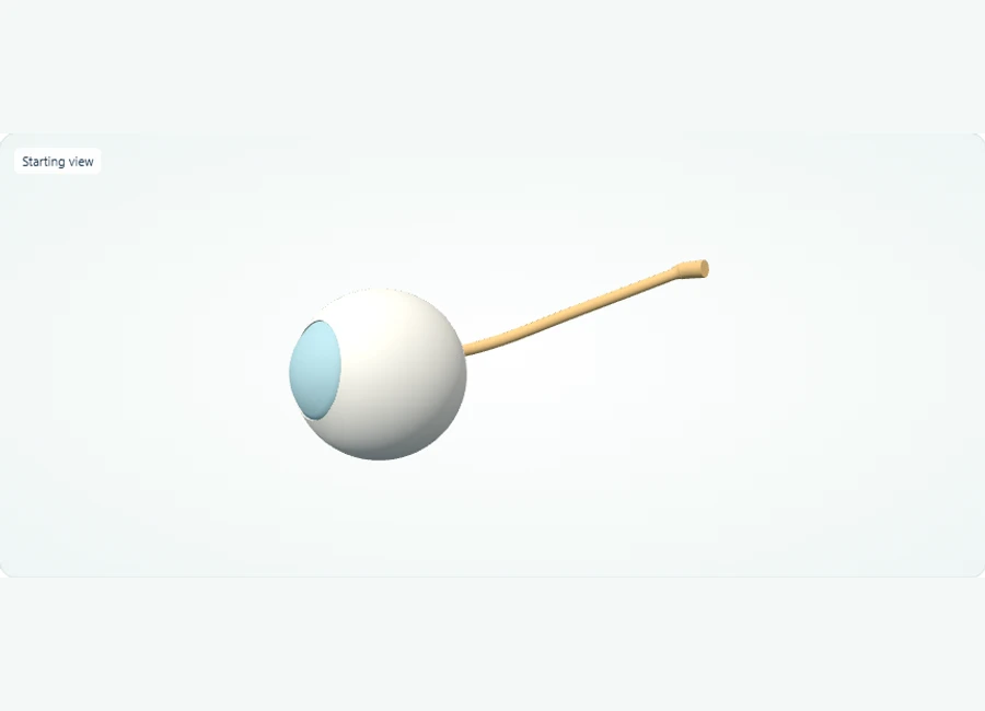
Rotate, isolate structures, and test your recall.
Select and isolate the right cornea, then the right lens, to inspect these anterior structures individually. The study model uses opaque surfaces.
Use Show all. Select and hide the sclera to expose deeper layers, then isolate the retina to inspect its shape.
Use Show all and choose Side or Back. Select the optic nerve to trace its exit from the posterior eye.
Try it yourself
Which structure exits the posterior eye, and which light-sensitive layer lines the posterior interior?
Show answer and explanation
The optic nerve (cranial nerve II) exits posteriorly; the retina lines the posterior interior.
From the back of the model, the nerve trunk is the structure leaving the eyeball, and the retinal lining sits within the posterior interior. Note the entire retina is shown as one part, and the macula is not separately labelled in this model.
- Refractive structures (cornea and lens) are anterior; the light-sensitive retina is posterior.
- The optic nerve (cranial nerve II) leaves the eye posteriorly, carrying visual information toward the brain.
- The sclera is the opaque outer coat; hide it to expose the cornea-lens axis and the internal retinal lining.
Applying Prentice's Rule to Decentered Lenses Instead of Memorizing the Formula
Prentice's rule states induced prism equals decentration in centimeters times lens dioptric power. The real skill is setting up the problem: identifying the decentration and getting the prism base direction correct.
The formula is P = cF, where c is the distance in centimeters between the visual axis and the lens optical center, and F is the lens power in diopters. Base direction is where errors concentrate. A plus lens behaves like prisms stacked base-to-base, so its prism base always points toward the optical center; a minus lens base points away. Worked example: a +4.00 D lens sits with its optical center 5 mm nasal to the pupil. The eye views through a point temporal to the optical center, so c = 0.5 cm and P = 2 prism diopters, base directed toward the optical center — that is, base-in. The common mistake is applying the minus-lens rule and calling it base-out.
Extended scenario: both lenses of a +4.00 D pair are decentred 5 mm nasal to the pupils after an incorrect pupillary distance measurement, inducing about 2 prism diopters base-in per eye. The plausible mistake is assuming the error merely blurs vision and cannot explain the patient's new binocular complaints. The better decision is quantifying the induced prism and predicting its effect on the alignment demand the patient's vergence system must meet. It matters because it explains why patients report symptoms with prescriptions that are nominally accurate, and it turns an arithmetic exercise into a prediction about binocular function.
Build fluency by drilling setup, not just computation. For any optics problem, first write down what quantity is the decentration, what is the power, which direction the base points, and what the induced prism does to the image or to binocular alignment. Checking those four statements catches the sign and direction errors that arithmetic practice alone does not, because arithmetic drills produce fast wrong answers just as easily as fast right ones.
Matching Ocular Drug Classes to Mechanisms When the First Choice Is Complicated
Glaucoma drug classes differ by mechanism: aqueous suppression, trabecular outflow enhancement, or uveoscleral outflow enhancement. Mechanism-based reasoning exposes systemic interactions that a memorized first-line list conceals.
Organize the classes by their mechanism link. Beta-blockers reduce aqueous humor production through beta-receptors in the ciliary epithelium. Prostaglandin analogs primarily increase uveoscleral outflow. Alpha-2 agonists reduce production and increase uveoscleral outflow. Carbonic anhydrase inhibitors reduce production. Because topically administered drugs can reach the systemic circulation — the nasolacrimal drainage pathway bypasses first-pass metabolism — the mechanism a drug uses ocularly can have systemic counterparts, which is the interaction surface these items can probe.
Worked scenario: a paper case presents a 68-year-old managed with a systemic beta-blocker for a cardiac rhythm problem, newly diagnosed with open-angle glaucoma. The plausible mistake is selecting a topical nonselective beta-blocker out of habit from a memorized list, despite the mechanism-level concern that both agents act on beta-receptors and the topical drug can be systemically absorbed. The better decision is reasoning from mechanism to a class with a different action, such as a prostaglandin analog that increases uveoscleral outflow. It matters because it demonstrates the chain running through pharmacology: real treatment decisions weigh more factors than this exercise shows, but the item-relevant reasoning hinges on the mechanism link rather than drug-name recall.
Convert your pharmacology notes into a mechanism map: one column for the drug class, one for the mechanism at the tissue level, one for the expected intraocular effect, and one for systemic considerations that follow from the same mechanism. Deriving the trade-name and generic list from that map is fast; the reverse process — memorizing names and hoping the mechanism follows — leaves you unable to reason when a complication, interaction, or side effect appears in a stem.
Separating Disc Edema Look-Alikes Using Discriminating Features
Papilledema, papillitis, and pseudopapilledema can all present as an elevated or swollen-appearing disc. Discriminating features — laterality, vision, pain, and disc appearance — turn a shared appearance into a differential decision.
Textbook presentations give you the discriminating chain links. Papilledema reflects raised intracranial pressure and is classically bilateral, with vision usually preserved early. Optic neuritis with papillitis is classically unilateral, with central vision loss and pain on eye movement. Pseudopapilledema describes an anomalously elevated disc appearance — for example, from optic disc drusen — without true edema, typically with stable findings and normal vision. The mistake to avoid is treating a swollen-appearing disc as one disease process and reaching for a single cause before asking the discriminating questions.
Force the differential by requiring at least two discriminating questions per scenario: Is it one eye or two? Is vision affected, and is there pain? Does the appearance reflect true edema or an anomalous disc? Drilling this way builds the habit of running the finding link backward through multiple chains simultaneously, which is exactly what a differential-diagnosis item demands. Use the table below as a template and reproduce equivalent tables for your own look-alike sets, such as red eye causes or optic neuropathy patterns.
A note on how to use the table: do not memorize the rows as isolated trivia. Attach each row to its mechanism — for example, pain on eye movement in optic neuritis relates to inflammation of the affected nerve, and preserved early vision in papilledema relates to pressure affecting the disc before the macula. Mechanism-anchored rows survive the exam better than rows recited from a chart, because a stem can present any row as the entry point.
| Feature | Papilledema | Optic neuritis (papillitis) | Pseudopapilledema |
|---|---|---|---|
| Laterality | Typically bilateral | Classically unilateral | Often bilateral, anomalous discs |
| Vision | Usually preserved early | Central vision loss common | Typically normal |
| Pain | Absent | Pain with eye movement is characteristic | Absent |
| Disc appearance | True edema with obscured margins | Disc edema with inflammatory signs | Elevated appearance without true edema, e.g., drusen |
A Chain-Writing Exercise That Exposes Gaps Before Test Day Does
Pick five core topics, write each one's mechanism chain from memory under a time limit, then score it against a rubric. The rubric converts vague familiarity into specific, observable readiness signals.
Set up the exercise: choose five representative topics, such as aqueous humor dynamics, the visual pathway, retinal phototransduction, one glaucoma drug class, and accommodation. For each topic, write in five minutes or less the three-link chain — normal function, disruption, predicted findings — plus one clinical question the chain answers. Then open a reference and mark every link that is missing, vague, or reversed in causal direction. The gap pattern across the five chains, not any single score, is the informative result.
Score each chain with the rubric below. Two expected observations tell you what to do next: a combined score below 21 out of 30 signals that reference review of the failing areas should come before more question practice, and any chain whose finding link simply restates the diagnosis — writing that glaucoma causes glaucomatous damage rather than naming the mechanism — marks a pharmacology or pathology gap specifically. Treat these as learning milestones for pacing your review, not as predictions of any score outcome.
Repeat the exercise weekly with a fresh set of five topics. Watch two trends over repetitions: chains should take less time to write cleanly, and the failure pattern should migrate from the normal-function link toward the finding link as your science base solidifies and your remaining work shifts to application. If failures stay clustered at the disruption link in one content area, that area needs its own focused chain-writing session rather than mixed review.
- 2 points: the link is specific, causal, and stated at the correct level — cellular, tissue, or systemic
- 1 point: the link is present but vague, or stated in the wrong causal direction
- 0 points: the link is missing, or the finding link merely restates the diagnosis
- Expected observation: a total below 21 of 30 across five chains means reference review precedes further question practice
- Expected observation: chains completed cleanly within five minutes indicate a topic ready for spaced review rather than new learning
An Adaptable Sequence With Concrete Readiness Checks
Sequence preparation in three phases: build chains per content area, then integrate across areas with mixed scenarios, then sharpen with timed case-style sets. Readiness checks, not calendar dates, control when you advance.
Phase one, roughly three weeks if you have a standard runway: build chains within each content area — anatomy and physiology together, then optics, pharmacology, and pathology — using the weekly exercise above. Phase two, about four weeks: integrate, using paper scenarios that require two chains at once, such as a drug mechanism applied to a pathway finding or an optical calculation embedded in a binocular-vision complaint. Phase three, about two weeks: timed case-style sets with an error log keyed to chain links rather than to right and wrong answers. Compress or stretch phases to fit your actual calendar.
Transition between phases based on the checks below, not the calendar. One administrative note: registration, policies, fees, scheduling, and accommodations for NBEO examinations are maintained by NBEO on its own exam pages — verify those specifics there rather than relying on secondary summaries, and keep your preparation plan focused on content reasoning, which is what this guide addresses.
The error log deserves its own discipline. For every missed practice item, record which chain link failed: did you misidentify the normal function, miss the mechanism, or fail to connect the mechanism to the finding? A log showing misses concentrated in one content area redirects your remaining time to that area; a log showing misses scattered randomly signals readiness to spend time on timed conditions instead. Both patterns are informative; an unlogged miss teaches you nothing for next time.
- You can write a complete three-link chain, unprompted, for every topic you flagged as high-yield
- For each missed scenario, you can name the exact chain link where your reasoning diverged, not just the correct answer
- Your optics setups consistently get base direction right before you worry about solving them quickly
- Your error log shows misses concentrated in identifiable content areas rather than scattered without pattern
References and further reading
Use these references to explore the concepts and check the latest information from the relevant organizations.