
Study for the CPSGT by pairing concepts that look alike and forcing a decision between them each time: obstructive versus central apnea, hypopnea versus RERA, N1 versus wake, arousal versus awakening, real slow waves versus sweat artifact. Work through scored examples, write the rule you applied, and log every ambiguous call as a review item.
Why respiratory event classification is a definitions problem, not a waveform problem
Apneas, hypopneas, and RERAs are distinguished by explicit definitional criteria, not by how dramatic a tracing looks. Train yourself to check each criterion in order before naming the event.
A flat-looking airflow channel can tempt you to call everything an apnea, while a modestly reduced but prolonged flow limitation pattern can be overlooked entirely. The corrective habit is a fixed sequence: measure the event duration, estimate the amplitude reduction in airflow, inspect the effort channels, and then look for an associated oxygen desaturation or arousal. Only after those observations do you assign the event type. Practicing this sequence on paper examples builds the discipline that flashcard recall alone does not.
The definitions used in sleep teaching also come in slightly different rule sets, so know which criteria your training program follows and state them explicitly in your notes. For example, the amplitude reduction and the desaturation-or-arousal requirement commonly attached to hypopnea scoring can differ between scoring frameworks. Write the version you are using at the top of your practice worksheet, then apply it consistently. When you review scored recordings later, disagreements about event type often turn out to be disagreements about which rule was applied, which is exactly the kind of confusion you want to eliminate before the exam.
- Decision sequence: duration first, then airflow reduction, then effort, then desaturation or arousal.
- Name the rule set you are applying before you score a single event.
- Treat an ambiguous event as a review item, not a guess to be forgotten.
| Event | Airflow pattern | Effort channels | Commonly attached requirements |
|---|---|---|---|
| Obstructive apnea | Markedly reduced or absent | Continued, often out of phase | Duration meets minimum; often paired with desaturation or arousal |
| Central apnea | Markedly reduced or absent | Reduced or absent | Duration meets minimum; no associated effort |
| Mixed apnea | Absent throughout | Absent early, resumes later | Effort pattern shows both phases |
| Hypopnea | Reduced but not absent | Usually continued | Amplitude reduction plus desaturation or arousal, per the rule set in use |
| RERA | Flattening or flow limitation | Continued | Leads to arousal without meeting apnea or hypopnea criteria |
Central versus obstructive apnea: what the effort belts actually decide
The effort channels, not the airflow channels, separate central from obstructive events. Continued chest and abdominal effort during an airflow pause indicates an obstructive pattern; absent effort indicates a central one.
Worked scenario: a practice excerpt shows twenty seconds with a flat nasal pressure channel and a flat thermal signal. A first-pass reader labels it a central apnea because the airflow is gone. The better decision is to look at the inductance belts before naming anything. The chest belt shows continuing deflection and the abdominal belt moves out of phase with it, which is the signature of obstructive physiology: the patient is working to breathe against a closed or narrowed airway. The correct classification here is obstructive, and the out-of-phase effort is the observation that justifies it.
Why it matters: the classification you assign is part of the technologist's record that the sleep physician reviews, and mixing up effort patterns changes the clinical picture the report conveys. On the tracing itself, a central event and an obstructive event can look identical in airflow alone. Build the reflex of reading the belts first. A useful drill is to cover the airflow channels, look only at effort, and predict the event type before uncovering the rest. If your prediction and the airflow pattern disagree, stop and write down which observation overrode the other and why.
Explore in 3D: Right eye: layers, lens, and optic nerve
This model shows the right eye as five selectable parts: cornea, lens, retina, sclera, and optic nerve. The cornea and lens sit anteriorly, the retina lines the posterior interior, and the optic nerve exits posteriorly. Surfaces are opaque until hidden, so hiding the sclera is the key step to see inside.
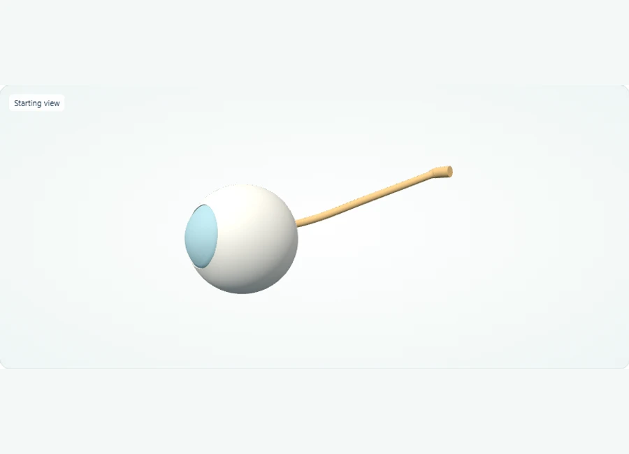
Rotate, isolate structures, and test your recall.
Select and isolate the right cornea, then the right lens, to inspect these anterior structures individually. The study model uses opaque surfaces.
Use Show all. Select and hide the sclera to expose deeper layers, then isolate the retina to inspect its shape.
Use Show all and choose Side or Back. Select the optic nerve to trace its exit from the posterior eye.
Try it yourself
Which structure exits the posterior eye, and which light-sensitive layer lines the posterior interior?
Show answer and explanation
The optic nerve (cranial nerve II) exits posteriorly; the retina lines the posterior interior.
From the back of the model, the nerve trunk is the structure leaving the eyeball, and the retinal lining sits within the posterior interior. Note the entire retina is shown as one part, and the macula is not separately labelled in this model.
- Refractive structures (cornea and lens) are anterior; the light-sensitive retina is posterior.
- The optic nerve (cranial nerve II) leaves the eye posteriorly, carrying visual information toward the brain.
- The sclera is the opaque outer coat; hide it to expose the cornea-lens axis and the internal retinal lining.
Stage scoring boundaries: N1 versus wake, and N2 versus N3
Stage boundaries are defined by specific traceable features: slowing and reduced alpha for N1, spindles or K-complexes for N2, and slow-wave proportion for N3. Learn the features as checklist items, not impressions.
N1 is the boundary stage that produces the most hesitation, because drowsy tracings drift in and out of alpha. Instead of judging overall look, anchor on named features: attenuation of alpha relative to wake, slower theta activity, slow eye movements, and changes in chin tone. If alpha is still dominant in the epoch, the call is wake regardless of how sleepy the rest of the tracing appears. Writing these anchors on a single card and checking each epoch against them converts a vague impression into a repeatable decision.
N2 versus N3 is a proportion problem. N2 is characterized by the presence of spindles or K-complexes on a background that does not meet the slow-wave threshold, while N3 is defined by a substantial proportion of slow-wave activity in the epoch. The common practice mistake is to let a single prominent slow wave push an epoch into N3, or to miss that slow waves occupy most of an epoch during a deep-sleep stretch. Drill this by scoring consecutive epochs across a transition and noting exactly which feature justified each stage change, so the boundary is traceable rather than felt.
- N1 anchors: reduced alpha, theta background, slow eye movements, falling chin tone.
- N2 anchors: sleep spindles and K-complexes on a non-slow-wave background.
- N3 anchor: slow waves occupying a large proportion of the epoch, per your training criteria.
- Traceability check: for any stage call, name the feature that made it.
Arousals and awakenings: two concepts a short duration separates
An arousal is a brief shift toward lighter sleep or wake with an acceleration in EEG frequency; an awakening means the epoch ends in wake. Duration and outcome distinguish them, and both must be tied to observable tracing changes.
The practical difficulty is that K-complexes, deltas, and sleep spindles are themselves bursts of faster or higher-amplitude activity, so not every frequency change is an arousal. Teaching criteria require the acceleration to last a minimum duration, be at least a few seconds, and follow defined rules about which sleep stage it occurs in and which channels show the change. When you score a candidate arousal, ask two questions: does the acceleration meet the duration minimum, and does the epoch end in wake? A no to the second question keeps it an arousal; a yes makes it an awakening.
Train this with a paired-exercise approach. Take a practice recording and mark every abrupt frequency change you can find, then sort the marks into three buckets: arousal, awakening, and normal sleep-graphic element such as a spindle or K-complex. The sorting is where understanding develops, because you must articulate why a three-second burst qualifies and a one-second burst does not. Record your sorting decisions alongside the definitions. If you later review a scored version of the same excerpt, your disagreements will point precisely at which definitional element you applied loosely.
Artifacts that imitate pathology: sweat, electrode pop, and ECG contamination
Artifacts are recurring channels-and-sources problems: sweat slows the EEG, a popped electrode produces sharp transients, and ECG contaminates nearby channels. Recognition comes from comparing a suspect channel against its neighbors.
Worked scenario: during a warm night, an EEG channel begins to sway with very slow, undulating deflections that a reader interprets as deep-sleep slow waves, and the night's stage summary shifts toward more N3. The better decision is a cross-channel check: sweat artifact characteristically appears on the channels near the affected area while relatively distant references look normal, and the undulation rhythm is slower than physiological slow-wave activity. Checking impedances, noting room conditions, and documenting the affected channels and time range is the appropriate technologist response, and it matters because uncorrected artifact distorts the stage percentages reported to the physician.
The same comparative habit handles other common contaminants. Electrode pop produces abrupt, repetitive sharp transients confined to one derivation and often follows a lead that has been disturbed; ECG artifact shows a rhythm locked to the cardiac channel, visible as periodic spikes in EEG and eye channels at the heart rate. For each suspect pattern, the disciplined questions are the same: which channels show it, is it time-locked to another physiological signal, and is the shape physiologically plausible? A paper exercise of labeling ten artifact examples by channel distribution, rather than by shape alone, cements this reasoning.
- Sweat artifact: slow swaying, regionally distributed, worse in warm conditions.
- Electrode pop: sharp transients confined to one derivation.
- ECG artifact: periodic contamination time-locked to the cardiac channel.
- Universal check: compare the suspect channel to neighboring references before interpreting.
Biocalibration and documentation: the record you leave behind
Before scoring begins, the recording must be verified channel by channel: signals present, impedances acceptable, each modality confirmed with an observed task. Documentation then captures what was checked, what changed, and what was done about it.
Biocalibration is a deliberate sequence in which the patient is asked to perform observable tasks while you confirm each modality responds correctly: eyes open and closed for the EEG and eye channels, biting or clenching for the chin EMG, breathing instructions for airflow and effort, and limb movements for the leg channels. The value of rehearsing this as a written checklist is that it mirrors the sequence of real lab practice and keeps the modality-by-modality topics in your study plan organized. A channel that was never confirmed is a channel whose later tracing you cannot fully trust.
Documentation is the companion skill. An event note should state what was observed, when, and what action was taken: a sensor was repositioned at a time stamp, an artifact was identified and its channels listed, a patient complaint was recorded. Contrast a strong entry with a weak one; 'artifact noted' is weak, while 'slow swaying artifact on left central and left occipital derivations from roughly the third hour, impedances rechecked, patient reported warmth' is usable by anyone reading the record later. Practicing writing entries in that structure is a low-cost exercise that reinforces the methods and documentation topics directly.
- Confirm every modality with an observable patient task during biocalibration.
- Document observations with channel names, time ranges, and actions taken.
- Treat an unverified channel as unreliable until it is checked.
A workable preparation sequence and a self-check rubric
Sequence your preparation in four passes: build paired-concept notes, score practice excerpts, drill artifact labeling, and finish with mixed scenarios under time. Measure readiness by rubric scores on each pass, not by hours studied.
A realistic adaptable sequence: first pass, one to two weeks writing contrast notes for each paired concept (apnea types, hypopnea versus RERA, arousal versus awakening, N2 versus N3, artifact versus physiology), stating the decision rule in one sentence. Second pass, score short practice excerpts and log every ambiguous call. Third pass, an artifact-only drill on labeled examples. Fourth pass, mixed case-style scenarios where several concepts arrive in a single case, which builds the integration that isolated topic drills cannot. Compress or stretch the passes to fit your schedule, but keep the ordering, because later passes depend on the contrasts built earlier.
Practical exercise with expected observations: take a scored ten-minute excerpt from your training materials or a public teaching recording, and before looking at any scoring annotations, mark respiratory events, stages, and arousals yourself. Expected observations on a first attempt are that you will under-call short hypopneas, hesitate on N1 epochs, and misread at least one artifact segment; the point of the exercise is the discrepancy log, not a perfect sheet. Self-check rubric, with scores as learning milestones rather than predictions of any exam result: two points for each event you can justify by naming the rule applied, two points for each artifact you identified by channel distribution, and two points for each stage call where you named the defining feature. Re-run the exercise weekly and watch the justification quality, not just the count, improve.
- Readiness check 1: you can state the decision rule for each paired concept from memory.
- Readiness check 2: your discrepancy log from practice excerpts is empty of repeats.
- Readiness check 3: you can write a complete documentation entry for an artifact and a sensor adjustment.
- Readiness check 4: in mixed scenarios, you complete the observation sequence before naming any event.
References and further reading
Use these references to explore the concepts and check the latest information from the relevant organizations.